Understanding heart anatomy, congenital defects, and disease variations is essential for treatment and education in pediatric cardiology. Traditionally, this relied on physical specimens, models, and 2D images. However, advances in 3D scanning have transformed the study and teaching of heart anatomy and pathology.
Professor Dr. Rohit Loomba, a pediatric cardiac physician at Lurie Children's Hospital in Chicago, has been at the forefront of this change. By using the Revopoit MIRACO 3D scanner, Dr. Loomba has contributed to research and medical education. This case study explores how 3D scanning has transformed the understanding and learning of pediatric cardiology.
Limited Access to Heart Specimens
One of the primary challenges in pediatric cardiology is the scarcity of anatomical heart specimens, particularly those with congenital malformations.
As Dr. Loomba explains, “There are not a large number of anatomic specimens available throughout the world, specifically hearts. There are even fewer hearts that have congenital malformations. Thus, a large number of people do not have access to heart specimens to help them learn about the anatomic nuances.”
The restricted access is further complicated by challenges in preserving physical specimens, especially those stored for extended durations or needing transportation. On top of that, traditional models like wax molds or plastic replicas often don't capture the full complexity and spatial relationships of heart structures. This can leave students and clinicians struggling to understand the intricate details of congenital heart issues and diseases.
3D Scanning to Bridge the Gap
3D scanning has proven to be an invaluable tool in overcoming these barriers. Using 3D models created with the Revopoint MIRACO, Dr. Loomba can provide a highly detailed, accurate, and accessible representation of heart anatomy, regardless of geographical location. As Dr. Loomba notes, “Being able to use 3D scanning to create 3D models helps make these hearts accessible to others as well. While they may not have the actual heart in their hands, they can turn the 3D models and zoom in and zoom out…to learn about the structures and their spatial relationships.”

The ability to manipulate these digital models offers a level of interaction that traditional two-dimensional drawings, photos, or even videos cannot match. Medical professionals and students can explore the heart from various angles, zoom into specific structures, and understand the spatial relationships between different anatomical components.
Dr. Loomba further highlights the potential of 3D scanning for broader educational applications, “I have some collaborators who are interested in other parts of the body and was able to scan a knee joint, which turned out beautifully. There is lots of potential.” This ability to scan not just hearts but other anatomical structures opens new avenues for learning across disciplines.
Enhanced Education and Remote Collaboration
One of the most significant benefits of these 3D models is medical education. Traditional methods, such as textbooks, static models, and even physical specimens, are limited by accuracy and availability. Dr. Loomba notes, “Why use plastic heart models or drawings when you can use a scan of an actual heart? This ensures that the spatial relationships are entirely realistic and that all structures that can be scanned are included.”
With his 3D models, Dr. Loomba can teach students, professionals, and collaborators remotely and at any time something previously not feasible. He continues, “I have used the scans for Zoom lectures, to supplement in-person lectures, and even to reinforce pertinent anatomic concepts during lectures at conferences.”
In addition, Dr. Loomba is working with an international team to develop a free educational tool called Cardioscape, which houses a range of 3D heart models, allowing for interactive learning and better engagement. This tool will be handy for early learners and experienced clinicians alike, providing access to a diverse range of models that would otherwise be inaccessible.
Advantages Over Traditional Anatomical Models
The interactive nature of 3D scans provides a level of flexibility that traditional models cannot match. As Dr. Loomba explains, “With 3D scans, all the spatial relationships are retained, and the scan can be visualized from whatever angle or zoom one prefers. No visible structures are left out for any reason...Specific anatomic structures can be labeled.”
This increased interactivity enhances the learning experience, allowing for a more comprehensive understanding of heart anatomy and pathology.

Improved Access and Preservation
Another advantage of digital 3D models is their accessibility. Physical specimens require significant care, maintenance, and storage.
Dr. Loomba, who also works with two of the largest heart specimen archives in the world, the Maurice Lev Congenital Heart and Conduction Center and the Farouk Idriss Cardiac Registry, is well aware of the challenges of maintaining physical heart archives:
“Maintaining an archive of hearts is difficult for several reasons. First, you need someone who has an interest in it and can dedicate time to it. The hearts must physically be maintained.” In contrast, digital models do not degrade over time and do not require the same level of expertise for preservation.
Dr. Loomba envisions a future where digital registries of heart models are established, making it easier to maintain and share valuable specimens without the risks associated with physical preservation, “Once we can establish digital registries, those will be much easier to maintain…no deterioration of specimens with time. 3D scanning will let us immortalize these specimens because physical specimens won’t last indefinitely.”
This digital shift will allow broader access to heart specimens, enabling more people to learn from them regardless of location.

Analyzing Variations in Heart Anatomy and Disease
3D scanning also facilitates the study of variations in heart anatomy, particularly for those with congenital malformations.
As Dr. Loomba explains, “3D scanning helps us study the variations in spatial relationships and structure formation across individuals. More importantly, it lets us highlight these to others.” By scanning the hearts of different individuals, including those with congenital conditions, Dr. Loomba and his collaborators can better understand how these anomalies manifest and compare them across different cases.
He mentions an upcoming study involving 3D models, “We are about to publish a study regarding the pulmonary valve in the setting of tetralogy of Fallot…We have some 3D models included in the upcoming paper!” This ability to share and manipulate models remotely allows for greater collaboration among researchers in different parts of the world.
How the MIRACO 3D Scanner Helped
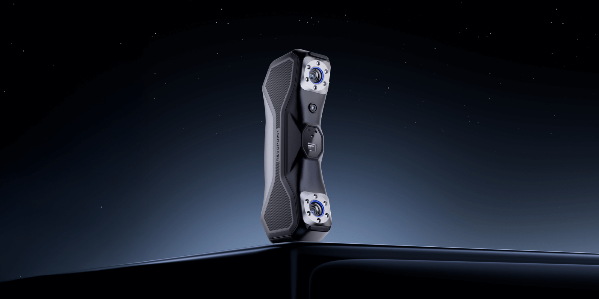
Dr. Loomba has found the Revopoint MIRACO scanner to be an ideal tool for his work due to its portability, affordability, and ease of use. Unlike the larger, more expensive scanners he first tried, the MIRACO is wireless and compact, allowing him to scan specimens at various archives without the burden of cumbersome equipment.
Dr. Loomba says, “The Revopoint MIRACO is portable, wireless, and affordable. I can put it into my backpack and have it with me if I am visiting other archives. It’s been easy to use, and the software has been intuitive!” This portability and ease of use enable Dr. Loomba to scan a variety of specimens without the logistical challenges posed by larger, more expensive equipment.
Streamlining the Workflow
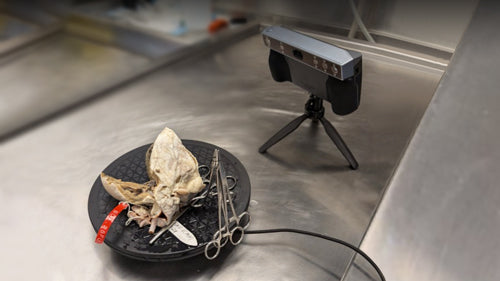
Dr. Loomba has developed an efficient workflow for scanning heart specimens. He selects hearts from the registry, prepares them, and positions them on a turntable using sutures and hemostats to expose key structures. Staging takes a few minutes to half an hour, depending on the specimen. Once positioned, Dr. Loomba uses the MIRACO to capture about 1,000 frames per scan.
After scanning, he uses Revopoint’s Revo Scan and Blender for post-processing. “I use platforms like Sketchfab and Cardioscape to house the models and make them accessible for teaching and collaboration,” he adds. This process enhances the learning experience and allows for the creation of a digital registry of heart specimens that can be shared globally.
A New Era in Pediatric Cardiology
3D scanning is transforming how heart anatomy and pathology are understood and taught in pediatric cardiology. Creating accurate, interactive models enhances education and collaboration and addresses specimen preservation challenges.
As Dr. Loomba’s work demonstrates, 3D scanning is not only an advancement in research tools but a paradigm shift in how medical professionals and students can learn and collaborate across disciplines.
Leave a comment
This site is protected by hCaptcha and the hCaptcha Privacy Policy and Terms of Service apply.